Topographical frequency dynamics within EEG and MEG sleep spindles
Summary
The study investigates how the frequency and location of sleep spindles change over their brief duration during stage 2 sleep. We found that spindles typically evolve from a faster, posterior-central pattern to a slower, anterior-frontal pattern, a dynamic that is clearly visible in EEG and present to a lesser extent in MEG.
Links
BibTeX tap to expand
@article{Dehghani_spindleTopo_2011,
title = {Topographical frequency dynamics within EEG and MEG sleep spindles},
journal = {Clinical Neurophysiology},
volume = {122},
number = {2},
pages = {229-235},
year = {2011},
issn = {1388-2457},
doi = {https://doi.org/10.1016/j.clinph.2010.06.018},
url = {https://www.sciencedirect.com/science/article/pii/S1388245710005432},
author = {Nima Dehghani and Sydney S. Cash and Eric Halgren},
keywords = {Spindle, Synchrony, MEG, EEG, Cortex, Thalamus, Inverse solution},
}
Code & Data
The room
Abstract
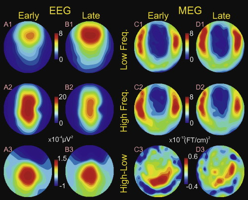
Spindles are rhythmic bursts of 10–16 Hz activity, lasting ∼1 s, occur during normal stage 2 sleep. Spindles are slower in frontal EEG and possibly MEG. The posterior-fast EEG pattern may predominate early in the spindle, and the anterior-slow pattern late. We aimed to determine the proportion of spindles showing this spatio-spectro-temporal interaction for EEG, and whether it occurs in MEG.
We recorded high density EEG and MEG from seven healthy subjects during normal stage 2 sleep. High vs. low frequency (12 vs. 14 Hz) power was measured early vs. late (25th–45th vs. 55th–75th duration percentile) in 183 spindle discharges.
The predicted spatio-spectro-temporal interaction was shown by 48% of EEG and 34% of MEG spindles (chance = 25%). Topographically, high frequency EEG power was greatest at midline central contacts, and low frequency power at midline frontal. This frequency-specific topography was fixed over the course of the spindle.
An evolution from posterior-fast to anterior-slow generators commonly occurs during spindles, and this is visible with EEG and to a lesser extent, MEG.
The spatio-spectral-temporal evolution of spindles may reflect their possible involvement in coordinating cortical activity during consolidation.
Citing
If you use this code or build on these ideas, please cite the paper using the BibTeX entry above.
Doors · concepts in this room
Related rooms
Evolutionary Optimization Reveals Structural Constraints on Reservoir Architecture for Spatiotemporal Chaos
Harnessing cortical geometry, wiring, and function as inductive biases for recurrent neural networks